")
")
Research Tools - Techniques
We use the following tools for our studies
1. Transient cerebral ischemia (stroke) via the filament Middle Cerebral Artery Occlusion (fMCAo) model.
We established in our Lab and routinely use the fMCAo model in mice. The model has been reviewed in detail elsewhere (Lourbopoulos et al, Journal of Cerebral Blood Flow Metabolism 2017, doi: 10.1177/0271678X16660986).
2. Complete and in depth neurobehavioral suite
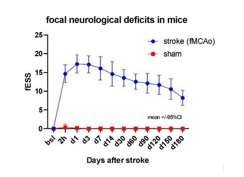
We developed and use a detailed neurological stroke scale, named Experimental Stroke Scale (ESS), graded from 0 up to 42 points of severity. The scale detects focal neurological deficits of a mouse after stroke and provides scores that are numerical - under restrictions and caution - similar to those of NIHSS used for human stroke. As an example, a score of 22 equals in both human and mice a severe and extensive hemispheric stroke. The ESS is a critical merging of all available and most used mouse stroke-behavioral scales up to date and has an excellent sensitivity and specificity for detecting neurological lesions in mice up to at least 6 months after stroke.
The scale can be found and downloaded here.
For detailed behavioral analysis we use the Noldus XT Ethovision Suite. <https://www.noldus.com/ethovision-xt>
We use the following behavioral tests, from acute up to long-term chronic phase after stroke:
• open field,
• novel recognition object,
• cylinder test,
• corner test,
• tail-suspension test,
• elevated plus maze,
• ladder rag walking test,
• adhesive-removal test.
Adaptively, we also develop or modify behavioral tests to serve the changes needs of our research.
3. Heart ultrasound of experimental animals after experimental stroke
4. Molecular analysis
We use established molecular techniques such as Western blot, PCR and Multiplex analysis, always depending on the relevant research question.
5. Microscopy analysis
We run multiple immunofluorescent analysis of brain and spinal cord tissue sections. We prepare fresh frozen or fixed crystat sections from our samples. We study our immunostained samples under a Leica Microscope, equipped with 4 fluorescent filters (dapi, FITC, TxRed, FarRed) and a monocrome FarRed-capable camera.
We recently upgraded our Microscope using customized 3D-printing techniques, in collaboration with the Technical University of Athens (Team and Laboratory of Prof. V. Spitas).
6. Cell cultures
We study in vitro macrophages, microglia and lymphocytes under different conditions afters stroke. We use molecular assays and immunofluorescence.